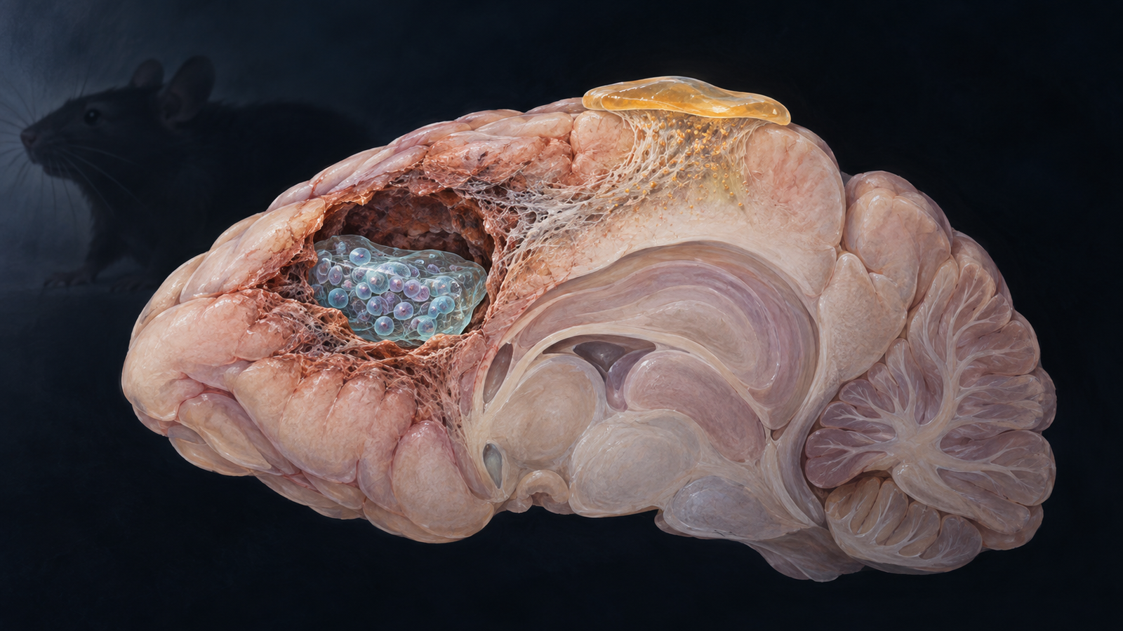
Editorial illustration of separate cavity and cortical-surface delivery systems used to combine a neural graft with sustained matrix modification. Credit: CellXperience generated editorial illustration.
Four weeks after human neural progenitors were placed into a rat stroke cavity, the investigators could no longer find them. The same cells remained detectable when a matrix-degrading enzyme had been delivered above the lesion from a second hydrogel.
The authors treated two different obstacles with two physically separate delivery systems: a hyaluronan-laminin gel supported the graft inside the cavity, while sustained-release ChASE37 modified the scarred extracellular matrix from the cortical surface.
The combination improved one gait measure and left surviving cells with early neuronal features. Yet grip strength improved after cells alone and after enzyme alone, too. Graft persistence, tissue markers, and motor recovery do not line up as a single causal chain. The study moves cell survival forward while leaving the source of functional recovery open.
Primary source
The complete peer-reviewed article was published online September 9, 2026. The full text, figures, methods, funding, and disclosure statement were available for review. The authors declare no conflicts and acknowledge a patent application for ChASE37.
The graft and the scar require different materials
An ischemic lesion can leave a fluid-filled cavity surrounded by reactive tissue. Neural progenitors injected into that space face mechanical stress, inflammation, poor access to trophic support, and an extracellular matrix that limits growth. A carrier can protect cells during and immediately after injection, but the neighboring scar remains a separate problem.
The team began by designing the cell carrier. Human iPSCs expressing GFP were differentiated into neural progenitor cells. Ketone- and aldehyde-modified hyaluronic acid were crosslinked to form a hyaluronan-oxime hydrogel, and laminin was added to support adhesion and neuronal differentiation.
A formulation containing 0.5% hyaluronic acid and 2 milligrams per milliliter laminin had a Young's modulus of about 0.40 kilopascals and supported a more even cell distribution than the softer formulation. A stiffer 1% hydrogel reduced survival and neurite length. The selected gel remained injectable through a 26-gauge needle and did not increase local microglial or astrocytic responses when tested in injured brain tissue.
ChASE37 could not share that carrier. The enzyme degrades hyaluronic acid, so mixing the components would dismantle the graft support. It was instead loaded into a chemically crosslinked methylcellulose hydrogel. Reversible binding within this depot was intended to extend enzyme availability.
The physical separation preserves a clear experimental question: can modification of the host extracellular matrix change what happens to a locally supported graft?
Five groups divide the contributions
The investigators created endothelin-1 lesions in the motor cortex and striatum of male Sprague-Dawley rats. Animals without a sufficient grip-strength deficit were excluded. The remaining rats were assigned at day four according to deficit severity so that groups began from similar functional values.
Treatment occurred seven days after injury. This subacute time point allowed a cavity and a maturing scar to form before a second procedure. The five groups received injury controls, both empty hydrogels, neural progenitors with an empty cortical depot, ChASE37 with an empty cavity gel, or the complete combination.
Each cell-treated animal received 50,000 neural progenitors in four microliters of hyaluronan-laminin hydrogel. The material was injected into the lesion at 0.5 microliters per minute. ChASE37 groups received 0.3 units of enzyme in six microliters of methylcellulose hydrogel placed epicortically above the injury. All experimental groups received tacrolimus, first by injection and then through an implanted pump.
Grip strength was followed to day 35 after injury. A handler blinded to treatment performed the assay. Gait was measured with the CatWalk system. Tissue was collected at day 35, four weeks after treatment, for scar, inflammation, lesion, graft, and cell-fate measurements.
This layout supplies controls for the injury, the delivery materials, each active component, and the combination. It cannot separate tacrolimus effects because immunosuppression was common to all groups, but that choice keeps rejection pressure more comparable across the xenograft experiment.
ChASE37 remained active without shrinking the lesion
Four weeks after one application, tissue in both ChASE37 groups contained degraded chondroitin sulfate proteoglycans. The groups without enzyme did not. This is direct evidence that the sustained-release formulation retained biological activity over the study window.
The tissue response was otherwise modest. Iba1-positive microglia and GFAP-positive astrocytes did not differ significantly among groups at the endpoint. Total lesion, infarct, and cavity volumes also did not differ significantly. Mean lesion measures were lower in the groups that received cells, but the paper presents that pattern as a suggestion rather than a demonstrated effect.
These results narrow the matrix claim. ChASE37 changed its intended substrate and did not add a detectable late inflammatory response under the measured conditions. The experiment does not show that it rebuilt lost tissue or reduced the established lesion.
An ex vivo result provides one possible connection to cell survival. Fresh brain slices from the injured hemisphere released more fibroblast growth factor 2 after ChASE37 exposure than after vehicle, about 3,749 versus 2,746 picograms per milliliter. The uninjured hemisphere had lower values and no significant treatment difference. Degrading the injury-enriched matrix may therefore release factors that had been bound within it.
That mechanism remains provisional. The slice experiment used three animals, and the in vivo study did not block FGF-2 to test whether it was necessary for graft persistence.
The surviving graft appears only with both components
At day 35, GFP-positive human cells were present in five of eight rats that had received the combination. No surviving graft was detected in the eight rats given cells without ChASE37. Human-specific nuclear and cytoplasmic antibodies supported the origin of the GFP-positive population.
The surviving cells extended processes into host tissue and expressed several early neural markers. Approximately 77% were positive for DCX, 76% for TUBB3, and 66% for GAP43. About half expressed GFAP, consistent with an astroglial component, while roughly 12% expressed NeuN. Pluripotency-marker OCT3/4 and proliferation-marker Ki67 signals were low but not absent.
Those percentages describe marker overlap within a small surviving population. They do not show mature neuronal identity. DCX and TUBB3 are compatible with migrating neuroblasts and immature neurons, and process extension is not evidence of synaptic connection. NeuN appeared in only a minority. Electrophysiology, host-to-graft tracing, graft-to-host tracing, and activity-dependent behavioral tests were not performed.
The four-week endpoint is also too early for a durable tumorigenicity or ectopic-differentiation claim. A small OCT3/4 or Ki67 fraction cannot establish long-term safety. It identifies a reason to extend observation and characterize the cells more deeply.
Motor recovery does not identify the working component
All injured rats began with an average grip-strength deficit of about 34% relative to their own pre-stroke baseline. After treatment, grip strength rose in the ChASE37-only, cell-only, and combination groups. By days 28 and 35, those groups approached the healthy controls and exceeded the injury-only and vehicle groups.
The broader gait assessment showed few group differences. At day 14, the combination improved the maximal contact area of the affected front paw relative to vehicle. That result agrees with better use of the stroke-affected forelimb, but it is one measure within a larger CatWalk panel.
Cells alone improved grip strength even though no graft was detected at the endpoint. Enzyme alone improved grip strength without donor cells. The complete combination left a graft, yet its grip-strength trajectory was not clearly superior to either component by itself. The authors also found no direct correlation between histological endpoints and functional outcomes.
A transient cell secretome could explain the cell-only behavior. Matrix digestion could assist host plasticity independently of transplantation. The combination may perform both jobs while adding long-term graft persistence. The present experiment cannot decide among these routes.
ChASE37 co-delivery was associated with detectable graft survival at four weeks, while the surviving graft's contribution to motor recovery remains unproven.
The delivery plan is modular and surgically demanding
The method has an appealing logic. Graft protection is tuned for the cells; matrix modification is tuned for the host tissue. Each material can be adjusted without forcing the other component into an incompatible formulation.
That modularity comes with a demanding procedure. Cells were injected inside the brain lesion, while a second hydrogel was placed on the cortical surface and held in contact with tissue. Translation would require a reproducible way to identify the cavity, deliver both materials without further injury, maintain depot position, control enzyme spread, and define an immunosuppression strategy for an allogeneic product.
Chondroitin sulfate proteoglycans also have context-dependent roles. Reducing an inhibitory matrix may support sprouting and migration, while excessive or poorly timed degradation could alter tissue boundaries and signaling. Dose, spatial confinement, release duration, and the post-stroke treatment window all become part of the product.
The next experiment should preserve the five-group design and lengthen follow-up. Aged animals of both sexes would better match the stroke population. Months of observation should track graft proliferation, migration, differentiation, and tumor risk. Electrophysiology and circuit tracing can test connection.
Selective graft silencing after mature connections form would reveal whether donor-cell activity is necessary for recovered movement.